Splenopancreatectomy, laparoscopic approach. Step-by-step surgery
Keywords:
hepatobiliary surgery, pancreatic surgery, malignant lesion of the distal pancreas, laparoscopic surgeryAbstract
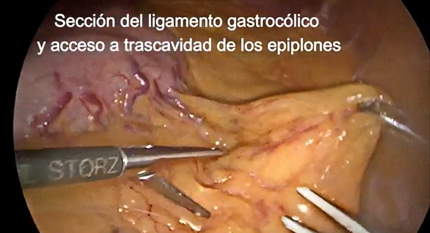
Distal pancreatic resections (to the left of the superior mesenteric artery), whether due to benign or malignant causes, represent a significant proportion of pancreatic surgeries.1,6 They are necessary procedures when curative treatment of a malignant lesion in the body and/or tail of the pancreas is indicated (often requiring splenectomy).2 Laparoscopic surgery, as is well known, offers several advantages over open laparotomy; this approach has evolved over recent decades, with studies validating its oncological safety.3,4 This procedure requires, as in all surgeries, an in-depth knowledge of regional anatomy and advanced surgical expertise to perform it safely both technically and oncologically.5,7A video is presented below of a 60-year-old female patient with a malignant-appearing lesion in the body and tail of the pancreas, identified on MRI and CT, which also revealed two lesions corresponding to simple cysts in the left liver lobe. A distal splenopancreatectomy was performed through a fully laparoscopic approach, detailing the step-by-step surgical procedure and subsequent postoperative course.
We highlight that, when defining the oncologic resection margin, it is considered positive (R1) when tumor cells are present less than 1 mm from the resection edge.8,9 A macroscopic margingreater than 1.5 cm from the resection border is therefore considered sufficient. In our patient's case, the lesion was located in the pancreatic tail, and the proximal transection was performed at the level of the suprapancreatic splenic artery, posterior to its origin from the celiac trunk, after it was divided between clips; therefore, the margin clearly exceeded the required distance.
The distal margin is also considered adequate, given that the spleen is included in the specimen. Finally, regarding lymph node dissection, peripancreatic nodes and those adjacent to the suprapancreatic splenic artery and splenic hilum corresponding to groups 10 and 11 of the Japanese surgical classification were removed.
Downloads
References
1.Evsen MS, Sak ME, Yalinkaya A, Firat U, Caca FN. A case of bifocal endometriosis involving a Pfannenstiel incision. Ginekol Pol. 2011;82(1):71-3.
2.Ozel L, Sagiroglu J, Unal A, Unal E, Gunes P, Baskent E, et al. Abdominal wall endometriosis in the cesarean section surgical scar: a potential diagnostic pitfall. J Obstet Gynaecol Res. 2012;38(3):526-30. doi: 10.1111/j.1447-0756.2011.01739.x.
3.Paramythiotis D, Karlafti E, Tsomidis I, Iraklis G, Malliou P, Karakatsanis A, et al. Abdominal wall endometriosis: a case report. Pan Afr Med J. 2022;41:193. doi: 10.11604/pamj.2022.41.193.33536.
4.Sahin L, Dinçel O, Türk BA. Rectus abdominal muscle endometriosis in a patient with cesarian scar: case report. Clin Exp Obstet Gynecol. 2013;40(4):599-600.
5.Kido A, Himoto Y, Moribata Y, Kurata Y, Nakamoto Y. MRI in the Diagnosis of Endometriosis and Related Diseases. Korean J Radiol. 2022;23(4):426-45. doi: 10.3348/kjr.2021.0405.
6.Gupta P, Gupta S. Scar Endometriosis: a Case Report with Literature Review. Acta Med Iran. 2015;53(12):793-5.
7.Sharmila V, Kamatham V, Shankaralingappa A. Abdominal scar endometriosis: A case report and review of literature. Indian J Pathol Microbiol. 2023;66(4):871-3. doi: 10.4103/ijpm.ijpm_460_22.
Downloads
Published
How to Cite
Issue
Section
License
Copyright (c) 2025 Alejandro Barboza, Alejandro Soumastre, Virginia Irigoyen, Gabriela Espinosa, Gabriel Massaferro

This work is licensed under a Creative Commons Attribution 4.0 International License.
All articles, videos and images published in Revista Cirugía del Uruguay are under the Creative Commons CC licenses, which is a complement to the traditional copyright, in the following terms: first, the authorship of the referred document must always be acknowledged and secondly none of the article or work published in the journal may have commercial purposes of any nature. The authors retain their copyrights and give the magazine the right of first publication of their work, which will be simultaneously subject to the Creative Commons Attribution-NonCommercial 4.0 International License license that allows the work to be shared whenever the initial publication is indicated in this journal.